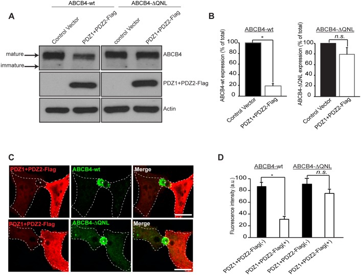
Fig 6. Effect of overexpression of the PDZ domains of EBP50.
(A) ABCB4-wt- or ABCB4-ΔQNL-expressing HepG2 cells were transiently transfected with the Flag-tagged EBP50 PDZ domains (PDZ1+PDZ2-Flag) or with the empty vector-Flag (control vector). ABCB4 was detected by immunoblotting from cell lysates. (B) Amounts of ABCB4 were quantified from immunoblots by densitometry. ABCB4 levels were expressed as a percentage of total expression in HepG2 cell transfected with control vector. (C) ABCB4-wt- or ABCB4-ΔQNL-expressing HepG2 cells were transiently transfected with the Flag-tagged EBP50 PDZ domains (PDZ1+PDZ2-Flag). Cells were fixed, permeabilized and stained with the anti-Flag antibody followed by anti-ABCB4 antibody and then incubated with Alexa-Fluor-594-and 488-conjugated secondary antibodies and visualized by confocal microscopy. Representative immunofluorescence images are shown. Asterisks indicate bile canaliculi. Bars, 10 μm. (D) The amount of ABCB4 at the bile canaliculi was quantified in HepG2 transfected cells (PDZ1+PDZ2-Flag(+)) and compared to that observed in control adjacent non-transfected cells (PDZ1+PDZ2-Flag(-)). Means (± SEM) of at least three independent experiments are shown. *P<0.01 for ABCB4-wt; n.s., not significant; a.u., arbitrary units.

