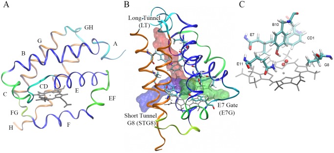
Fig 1. The distinctive truncated hemoglobin structure.
(A) Typical fold of a trHb structure and the commonly used structural blocks (shown in different colors). (B) Schematic representation showing the three different ligand entry tunnels present in trHbs: Long Tunnel (in red), Short Tunnel G8 (in blue) and E7 Gate (in green). (C) Schematic representation of the five structural positions that define the active site in a typical trHb. The figure shows the heme group (grey), the bound oxygen ligand (red in balls and sticks) and the five key residues (shown as sticks).

