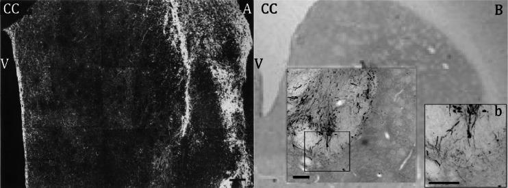
Fig. 2.
Comparison of grafted human foetal VM tissue and hiPS cell-derived DA neurons. a Overview of TH+ neurites in a coronal section of the striatum 4 months after grafting human foetal VM tissue from a 10-week-old foetus. The graft, filled with TH+ cell bodies and fibres is seen at the far right, TH+ processes radiate into host striatum and form a network that covers the whole area of striatum. b Overview of TH+ iPS cell-derived DA neurons (unsorted) 4 months after intrastriatal transplantation. Higher magnification showing TH-positive somata and some innervation of the surrounding host striatum by graft-derived neurites (b) Scale bar = 200 μm, CC = corpus callosum, V = ventricle. Adapted from [40, 75]

