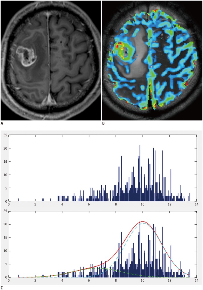
Fig. 2. Example of group 3 patient with negative skewness and leptokurtosis.
A. 54-year-old man with pathologically proven glioblastoma. Contrast-enhanced T1-weighted image showing contrast-enhancing mass in right frontal lobe. B. MR perfusion image showing increased nCBV in corresponding contrast-enhancing-lesion. C. Histogram derived from nCBV had negative skewness and leptokurtosis (skewness = -0.76, kurtosis = 3.52). nCBV = normalized cerebral blood volume

