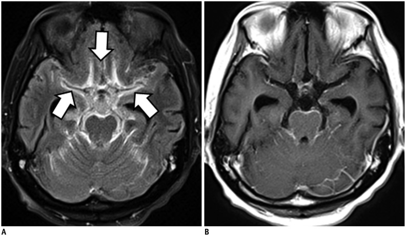
Fig. 6. Bithalamic glioblastoma with extensive CSF dissemination.
CE-FLAIR imaging (A) depicts more definite leptomeningeal enhancement (arrows) than CE-T1WI (B). Bithalamic inhomogeneous enhancing masses are noted and confirmed histologically as glioblastoma (not shown). CE = contrast-enhanced, FLAIR = fluid-attenuated inversion recovery, T1WI = T1-weighted imaging

