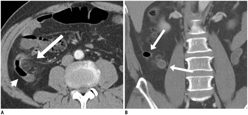
Fig. 2. 49-year-old male patient diagnosed as acute appendicitis by surgery.
A, B. Contrast-enhanced transverse (A) and coronal (B) CT images show characteristic findings of acute appendicitis, including appendiceal enlargement, appendiceal wall thickening and enhancement, and periappendiceal fat stranding. Appendix (arrows in A and B) is abnormally enlarged and dilated with air-fluid level in lumen (arrowhead in A).

