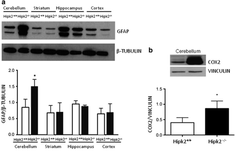
Figure 6.
Lack of HIPK2 induces a strong increase in astroglial cells. (a) Western blot analysis performed using GFAP antibodies of proteins extracted from cerebellum, striatum, hippocampus, and cortex. One representative experiment is shown. β-Tubulin was used for normalization. Densitometric analysis of three independent experiments is shown. Student's t-test, n=3 for each genotype. Data represent the mean±S.D. *P<0.05. (b) Western blot analysis performed with total extracts from the cerebellum of wild-type and Hipk2−/− mice using COX2 antibody. Vinculin was used for normalization. One representative experiment is shown. Densitometric analysis of western blot was performed using COX2 antibodies. For this analysis, four samples of wild-type mice and four samples of Hipk2−/− were used. For statistical analysis, Student's t-test was used for each genotype. Data represent the mean±S.D. *P<0.05

