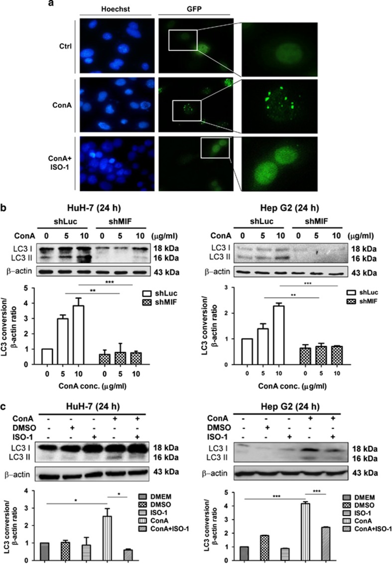
Figure 5.
MIF participates in ConA-induced autophagy of human hepatoma cells. (a) HuH-7 cells transfected with pEGF-LC3 plasmid were treated with ConA (20 μg/ml) in the presence or in the absence of ISO-1 (50 μM) for 24 h. The images were taken by fluorescence microscopy at a magnification of × 400 and × 600. Hoechst and GFP represents the nucleus and LC3 punctae, respectively. (b) HuH-7-shLuc/shMIF and Hep G2-shLuc/shMIF cells were treated with ConA at different doses as indicated for 24 h. Cell lysates were subjected to detect LC3 conversion by western blotting. (c) HuH-7 and Hep G2 cells were treated with ConA (20 μg/ml) in the presence or in the absence of ISO-1 (50 μM) for 24 h. Cell lysates were subjected to detect LC3 conversion by western blotting. Quantification of LC3 conversion is presented as the fold increase of LC3-II compared with the control cells after normalization to the corresponding β-actin levels. All data are presented as the mean±S.D. from at least triplicate independent experiments; *P<0.05, **P<0.01, ***P<0.001, ns indicates no significance

