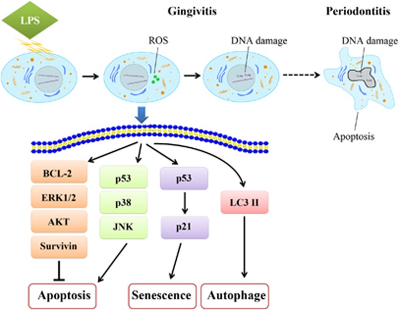
Figure 1.
Mechanisms of gingival fibroblasts in cell apoptosis resistance. LPS-induced ROS accumulation in acute inflammation. DNA damage appeared as time went on. Finally, apoptosis was detectable in chronic periodontitis. Short after stimulation, LPS increased phosphorylated ERK1/2, phosphorylated p38, phosphorylated JNK, p53, BCL-2, and Survivin in a dose-dependent manner. Simultaneously, phosphorylated AKT decreases in a dose-dependent manner. As a result, the anti-apoptotic effects and pro-apoptotic effects were balanced. The expression of p53 and p21, the ratio of LC3 ІI/LC3 I were increased by LPS, indicating senescence and autophagy were involved, respectively

