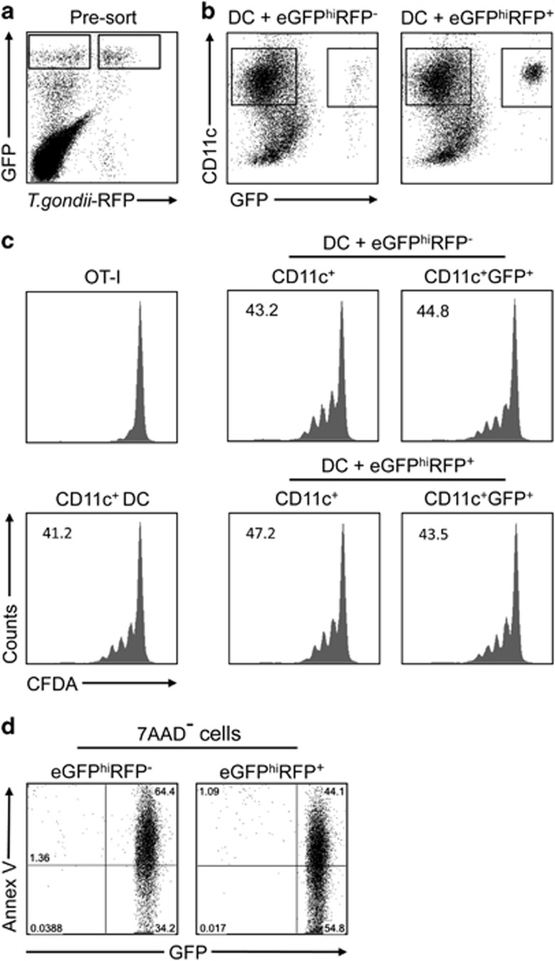
Figure 4.
Antigen presentation function of DCs that have captured T. gondii-infected and uninfected neutrophils. (a) eGFPhiRFP−-uninfected and eGFPhiRFP+-infected neutrophils recovered from the ear dermis 12 h after infection with 1 × 10⁶ T. gondii-RFP. (b) Representative dot plots of DCs cultured or not with eGFPhiRFP−-uninfected and eGFPhiRFP+ T. gondii-infected dermal neutrophils for 12 h and analyzed for expression of CD11c and GFP signal. Gates represent populations of CD11c+GFP− and CD11c+GFP+ cells. (c) Sorted CD11c+, CD11c+GFP− and CD11c+GFP+ cells were cultured with CFDA-labeled OT-I CD8+ T cells for 3 days in the presence of OVA antigen. Representative histogram plots of CFDA fluorescence of CD8+CD3+ gated cells showing the proliferative response. Numbers represent the frequency of cells with reduced CFDA content. (d) Representative dot plots of sorted eGFPhiRFP−-uninfected and eGFPhiRFP+-infected neutrophils recovered from the ear dermis 12 h after infection with 1 × 10⁶ T. gondii-RFP parasites and stained with annexin V-APC after gating on 7-AAD− cells. Quadrant values show the percentage of total gated cells. The data are representative of two independent experiments

