Figure 2.
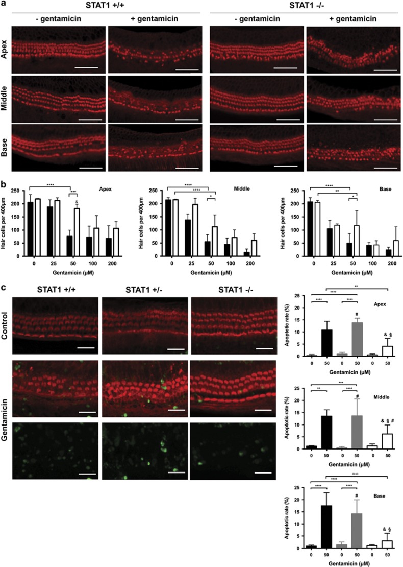
Gentamicin-induced hair cell damage was attenuated in STAT1−/− mice. (a) Representative confocal images of phalloidin-stained hair cells from all cochlear turns treated with 50 μM gentamicin for 24 h. At this gentamicin concentration, hair cell damage differed significantly between wild-type and STAT1−/− mice at all the cochlear turns. Gentamicin induced more hair cell damage in STAT1+/+ than in STAT1−/− mice. Scale bar for all figures, 50 μm. (b) Quantification of hair cell survival in wild-type (black) and homozygous (white) STAT1 mice. Explants were treated with different concentrations of gentamicin for 24 h. n=3–5 explants of each genotype and each condition. (c) Representative confocal images of TUNEL and phalloidin double-stained hair cells (middle turn) and quantification of apoptotic rate in wild-type (black), heterozygous (gray), and homozygous (white) STAT1 mice. Explants were treated with 50 μM gentamicin for 24 h. n=4–6 explants of each genotype and each condition. Scale bar for all figures, 20 μm. Values shown are mean+S.D. ****P<0.0001, ***P<0.001, **P<0.01, *P<0.05; &, not significant to control of same genotype; §, not significant to non-treated WT sample; #, not significant to gentamicin-treated WT sample

