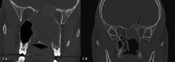
Fig. 5.

(A) Pre-operative coronal CT of case #10 who underwent a subtotal removal of the lesion with an endoscopic approach. (B) Coronal CT scan performed 6 months after surgery showing rapid regrowth of the remnant (arrow). An external approach was subsequently performed.
