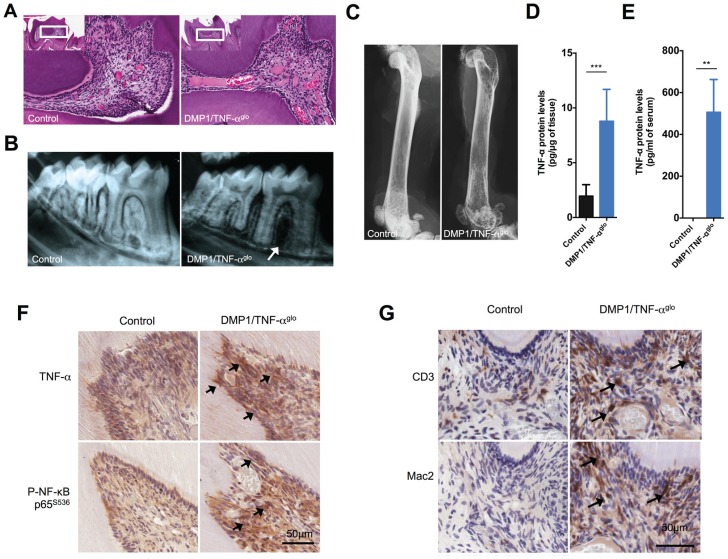
Figure 3.
Dentin matrix protein 1 (DMP1)/tumor necrosis factor–αglo (TNF-αglo) mice show inflammatory infiltrates within the tooth pulp. (A) Hematoxylin and eosin staining of the tooth pulp shows inflammation that is similar to pulpitis with infiltrating cells and enlarged blood vessels. (B) Radiographs also show alveolar bone loss around the teeth of the DMP1/TNF-αglo mice. (C) Femurs from the DMP1/TNF-αglo mice display reduced opacity, suggesting overall systemic bone loss due to overexpression of TNF-α by osteocytes. (D) Increased amounts of TNF-α were detected per µg of mandibular protein within the DMP1/TNF-αglo mice. (E) Serum levels of TNF-α were also increased in the DMP1/TNF-αglo mice as detected by a mouse TNF-α enzyme-linked immunosorbent assay (**P ≤ 0.01). (F) Immunohistochemical staining shows overexpression of TNF-α in the tooth pulp. Increased TNF-α cell signaling was also detected via phospho–NF-κB. Arrows denote sites of increased TNF-α expression and subsequent cell signaling via phosphorylation of NF-κB. (G) CD3 and Mac2 staining shows recruitment of lymphocytes and macrophages, respectively, in the tooth pulp. Arrows identify infiltrating lymphocytes and macrophages.

