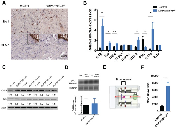
Figure 4.
Dentin matrix protein 1 (DMP1)/tumor necrosis factor–αglo (TNF-αglo) mice exhibit orofacial pain due to the TNF-α–induced pulpitis and osteitis. (A) Inflammation initiated by TNF-α is primarily restricted to the tooth pulp and bone and does not cause neuronal injury or glial activation within the trigeminal ganglia (TG) as determined by Iba1 and GFAP staining (lower inserts positive staining for central root astrocytes). (B) The expression of proinflammatory markers within the TG was evaluated by quantitative real-time polymerase chain reaction (*P ≤ 0.05; **P ≤ 0.01). (C) Cdk5 is a protein kinase; its activity can be upregulated by inflammation, so levels of both Cdk5 and its activator p35 were examined within the TG to determine if chronic tooth inflammation increased their expression. In most cases, the levels of both Cdk5 and p35 were higher in the DMP1/TNF-αglo mice versus the littermate control (fold differences were measured as the ratio over β-actin). (D) A Cdk5 kinase assay was performed using protein from the TG. Graph and representative blot showing modest increase in Cdk5 kinase activity. (E) Schematic of the dolognawmeter (Dolan et al. 2010), an assay and device used to quantify nociception by measuring masticatory function. (F) DMP1/TNF-αglo mice require more time to gnaw through a hard dowel compared to Cre− controls, signifying masticatory dysfunction and probable orofacial pain (****P ≤ 0.0001).

