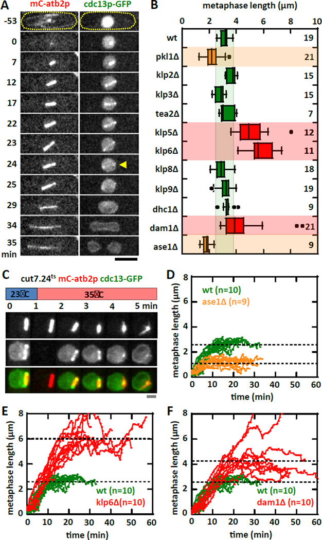
Figure 1. Motors and MAPs contribute to metaphase spindle length force-balance mechanism.
(A) Time-lapse images of a wildtype cell expressing mCherry-atb2p (tubulin) and cdc13p-GFP (cyclin) through mitosis. cdc13p is degraded from the spindle at the metaphase to anaphase transition (yellow arrow), marking precisely the final metaphase spindle length. The cdc13p-GFP marker is used in the screen for motors and MAPs affecting metaphase spindle length (see Fig. 1B). Bar, 5 µm.
(B) Targeted screen of fission yeast motors and selective MAPs for defects in metaphase spindle length at room temperature (23°C). Box plot shows spindle lengths - wildtype (3.1 ± 0.3 µm), pkl1Δ (2.0 ± 0.4 µm, p<10−4), klp2Δ (3.6 ± 0.3 µm, p<10−4), klp3Δ (2.8 ± 0.4 µm, p=0.1), tea2Δ (3.3 ± 0.5 µm, p=0.3), klp5Δ (5.3 ± 1.2 µm, p<10−4), klp6Δ (6.3 ± 1.6 µm, p<10−4), klp8Δ (2.9 ± 0.5 µm, p=0.4), klp9Δ (3.4 ± 0.4 µm, p=0.6), dhcΔ (3.4 ± 0.4 µm, p=0.1), dam1Δ (4.4 ± 1.6 µm, p<10−2), and ase1Δ (1.8 ± 0.3 µm, p<10−7).
(C) Temperature shift experiment of kinesin-5 cut7.24ts cells expressing mCherry-atb2p and cdc13p-GFP. Within 1 min of shifting to the non-permissive temperature of 35°C, the metaphase spindle exhibits spindle shortening and collapse, ultimately becoming a monopolar spindle. Note: The blank image at time 1-min in the cdc13p-GFP channel was due to thermal expansion of the coverslip causing an out-of-focus image, which was corrected in subsequent frames. Bar, 1 µm.
(D) Comparative plot of spindle length versus time of wildtype (green) and ase1Δ (organe) cells. Shown are pole-to-pole distances measured from prophase to the metaphase-anaphase transition. Wildtype metaphase spindles plateau at ~3 µm length. In contrast, ase1Δ metaphase spindles plateau at ~2 µm length.
(E) Comparative plot of spindle length versus time of wildtype (green) and klp6Δ (red) cells. In contrast to wildtype, klp6Δ metaphase spindles plateau at ~6 µm length.
(F) Comparative plot of spindle length versus time of wildtype (green) and dam1Δ (red) cells. In contrast to wildtype, dam1Δ metaphase spindles plateau at ~4 µm length.

