
Fig. 1.
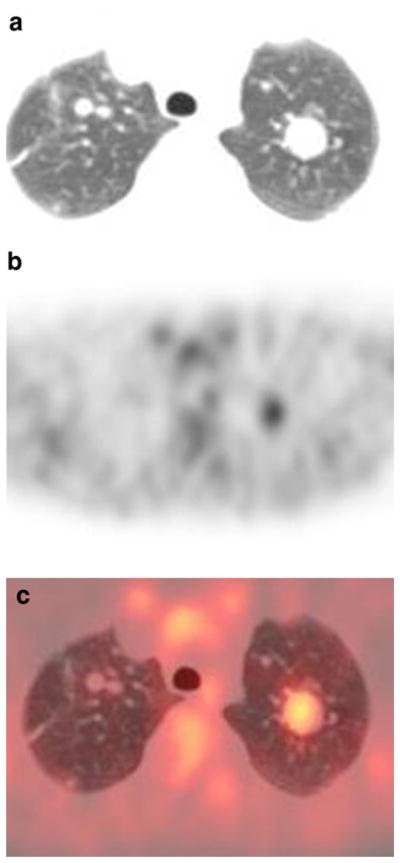
Patient 4: 9-year-old girl with bilateral lung metastases. a Transverse CT image of the lungs (lung window). b FDG PET at same level as a. c Fusion image of a and b. The largest lesion in the left upper lobe shows uptake higher than background lung. Smaller pulmonary lesions in the right upper lobe are not evident on the PET scan