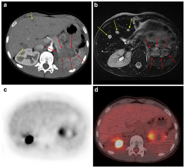
Fig. 2.
Patient 7: 10-year-old girl with left-sided recurrent Wilms’ tumor showing two liver metastases (yellow arrows) on contrast-enhanced CT (a) and three foci on MRI (b) not detectable on PET scan (c) or on PET/CT fusion image (d without intravenous contrast). The lesion in the pancreatic tail area (red arrows on a and b) is evident in all images

