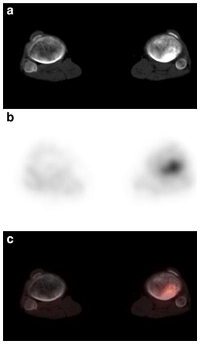
Fig. 4.
Patient 17: 11-year-old girl with recurrent Wilms’ tumor showing focal uptake and sclerotic process in the left proximal tibia; consistent with bony metastatic disease. a Noncontrast transverse CT image of the distal lower extremities (bone window). b FDG PET at same level as a. c Fusion image of a and b

