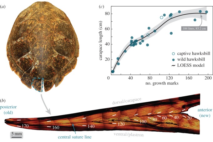
Figure 1.
Interior structure of the posterior marginal scutes of hawksbill sea turtles contains an extensive chronology. (a) Adult female carapace measuring 83.2 cm straight length. Blue dashed outline is the PM scute, where the largest tissue record on the carapace resides. White-dashed line is the cross-section path. (b) Polished, composite image of the longitudinal cross-section of the PM scute identifying major features. Growth trajectory (old to new) runs left to right. The dark suture line that runs horizontally separates the dorsal/carapace and ventral/plastron portions of the shell. Parallel records occur on either side of this line. Shadows at individual image edges are peripheral halos from microscope field-of-view. Scute coloration varies naturally. (c) Growth line count for 36 hawksbills, obtained from the PM scute. A LOESS model fit through the data shows an expected growth curve form with three apparent stages (0–45 cm, 45–75 cm and greater than 75 cm) corresponding to known hawksbill stage classes. Shaded area is the 95% confidence band.

