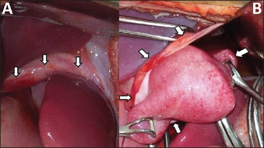
Figure 2.

(a) Defect in the diaphragm is seen with well-defined anterior lip (arrows). (b) Intraoperative image of the mass seen to occupy the cranial aspect of the diaphragmatic hernia sac (cut edges indicated by arrows)

(a) Defect in the diaphragm is seen with well-defined anterior lip (arrows). (b) Intraoperative image of the mass seen to occupy the cranial aspect of the diaphragmatic hernia sac (cut edges indicated by arrows)