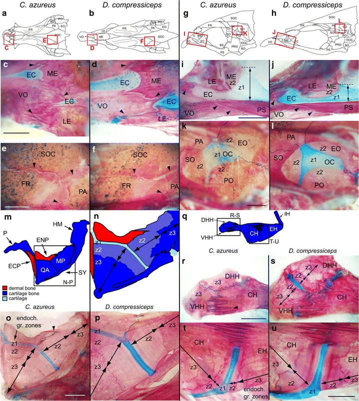
Fig. 10.

Shape and size differences in skeletal growth zones between CA and DC. a, b Camera lucida drawings of the dorsal aspect of CA (a) and DC (b) neurocrania with boxed areas highlighting suture regions shown in (c–f). c, d Dorso-lateral aspect of preorbital region showing sutures (arrowheads) between VO, LE and ME bones in CA and DC. e, f Dorso-lateral aspect of cranial vault region showing sutures (arrowheads) between FR, PA and SOC bones in CA and DC. Scale bar 100 μm. g, h Camera lucida drawings of the lateral aspect of CA (g) and DC (h) neurocrania with boxed areas highlighting suture regions shown in i–l. i, j Lateral aspect of preorbital region showing EC and sutures (arrowheads) between VO, LE and PS bones, and endochondral growth zone between EC and ME in CA and DC. Z1–2 zone 1–2 (k, l) dorso-lateral aspect of otic region showing sutures (lines) between EO, PO, SO and PA, and endochondral growth zone between OC and EO, PO and SO in CA and DC. Scale bar 100 μm. Z1–2 zone 1–2 (m, n) Camera lucida drawings of the suspensorium in CA (m) and endochondral growth zone between QA and MP (n). o, p Endochondral growth zone between QA and MP in CA (o) and DC (p). Scale bar 200 μm. Z1–3 zone 1–3 (q) Camera Lucida drawing of the ceratohyal complex skeleton in CA with boxed areas highlighting endochondral growth zones shown in r–u. r, s Endochondral growth zones and sutures (lines) between CH, DHH and VHH in CA (r) and DC (s). t, u Endochondral growth zones and sutures (lines) between CH and EH in CA (t) and DC (u). Scale bar 100 μm. Z1–3 zone 1–3. Bone nomenclature after [64]. DHH dorsal hypohyal, EC ethmoid cartilage, ECP ectopterygoid, ENP entopterygoid, EO epiotic, FR frontal, HM hyomandibular, LE lateral ethmoid, ME mesethmoid, MP metapterygoid, OC otic cartilage, P palatine, PS parasphenoid, PA parietal, PO pterotic, QA quadrate, SO sphenotic, SOC supraoccipital crest, SY symplectic, VHH ventral hypohyal, VO vomer
