
Fig. 14.
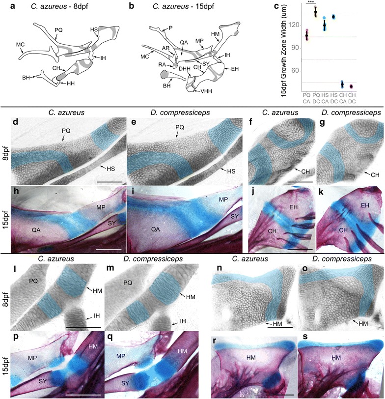
Development of endochondral growth zones in the pharyngeal skeletons of CA and DC larvae. a, b Camera lucida drawings of first and second pharyngeal arch cartilages in 8 and 15 dpf CA larvae showing endochondral growth zones (gray). a CA larval cartilages at 8 dpf. b Cartilage bones differentiating at 15 dpf from cartilage templates. c Mean and distribution of growth zone width in CA and DC in 15 dpf PQ, HS and CH (n = 5, each). d–s Higher magnification images of developing endochondral growth zones in dissected and flat-mounted alizarin red-/alcian blue-stained first and second pharyngeal arch cartilages in 8 dpf (blue shading d–g, l–o), and 15 dpf (h–k, p–s) CA and DC larvae. Scale bar 100 um. ***p < 0.001, Tukey’s HSD test. Bone nomenclature after [64]. AR articular, BH basihyal, CH ceratohyal, DHH dorsal hypohyal, EH epihyal, HH hypohyal, HM hyomandibular, HS hyosymplectic, IH interhyal, MC Meckel’s, MP metapterygoid, MX maxilla, P palatine, PQ palatoquadrate, QA quadrate, RA retroarticular, SY symplectic, VHH ventral hypohyal