Fig. 6.

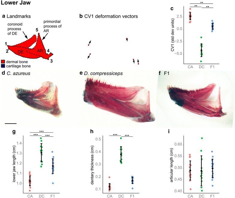
Shape and size differences between the lower jaw skeletons of CA, DC and F1 hybrids. a Camera Lucida drawing of CA lower jaw indicating anatomical landmarks. Dotted lines indicate approximate locations of bone boundaries. b CV1 deformation vectors on CA landmark configuration. c Mean and distribution of CA (n = 15), DC (n = 12) and F1 (n = 16) individuals along the CV1 axis, expressed in within-species standard deviation units. **p < 0.001, Bartlett’s test. d–f Dissected, alizarin red-/alcian blue-stained lower jaw skeletons of CA (d), DC (e) and F1 (f) specimens. Scale bar 2.5 mm. g–i Mean and distribution of CA, DC and F1 lower jaw length (g), dentary thickness (h) and articular process length (i). ***p < 0.001, Tukey’s HSD test. Bone nomenclature after [64]. AR articular, DE dentary
