Fig. 7.

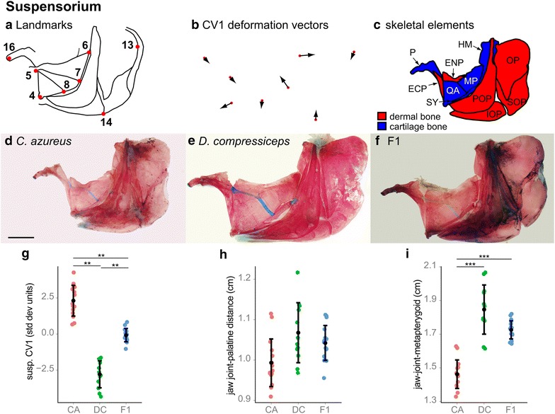
Shape and size differences between the suspensorium and opercular series skeletons of CA, DC and F1 hybrids. a Camera Lucida drawing of CA suspensorium and opercular series indicating anatomical landmarks. b CV1 deformation vectors on CA landmark configuration. c Camera Lucida drawing of CA suspensorium and opercular series bones. d–f Dissected, alizarin red-/alcian blue-stained suspensorium and opercular series skeletons of CA (d), DC (e) and F1 (f) specimens. Scale bar 5 mm. g Mean and distribution of CA (n = 15), DC (n = 12) and F1 (n = 16) along the CV1 axis, expressed in within-species standard deviation units. **p < 0.001, Bartlett’s test. h, i Mean and distribution of CA (n = 15), DC (n = 12) and F1 (n = 16) jaw joint-palatine distance (h) and jaw joint-metapterygoid distance (i). ***p < 0.001, Tukey’s HSD test. Bone nomenclature after [64]. ECP ectopterygoid, ENP entopterygoid, HM hyomandibular, IOP interoperculum, MP metapterygoid, OP operculum, P palatine, POP preoperculum, QA quadrate, SOP suboperculum, SY symplectic
