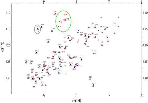
Fig. 5.
HSQC spectra of the isolated dSH3 domain (blue) and dSH3-ll-dSH3 tandem (red). The dSH3 resonances are labeled except for Trp, Asn, and Gln side-chain correlations. The elliptical contours outline the resonances from the glycine-rich linker and from the residue V9, which demonstrates peak doubling effect.

