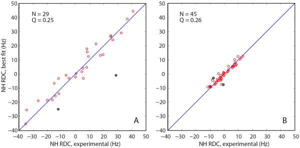
Fig. 6.
Fits of the experimental RDCs from (a) the isolated dSH3 domain and (b) dSH3-ll-dSH3 tandem with the structural model based on the coordinate set 1U06 (protonated, energy minimized). The fitting is limited to the core portion of the protein, residues 9–60, and does not include the data from N47 and D48 (grey circles in the plot). The total number of the fitted RDCs and the figure of merit Q are indicated in the graphs.

