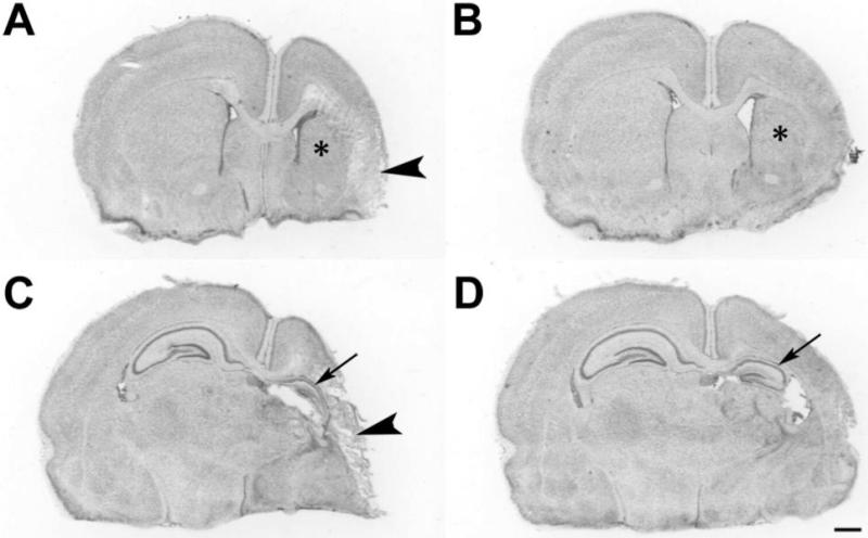
Figure 3. Neuropathology.
These cresyl violet stained coronal sections at the level of striatum (A,B) and hippocampus (C,D) illustrate representative histopathology in DHA+hypothermia (B,D) and albumin-hypothermia (A,C) treated animals on P14. After right carotid ligation+90 min 8% O2 exposure, P7 pups received DHA, 2.5 mg/kg (B,D) or 25% albumin vehicle, followed 1h later by hypothermia (3h, 30 °C). In the control (A,C) there is right cortical infarction (arrowheads), striatal atrophy and infarction (*) and hippocampal atrophy (arrow). In the DHA+Hypothermia brain (B,D), there is less right hemisphere, striatal (*) and hippocampal (arrow) atrophy (scale bar=1mm).

