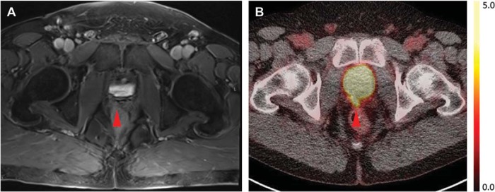
Figure 2.
(A) Axial T1-weighted postcontrast MRI and (B) axial [18F]DCFPyL PET/CT images through the prostate bed demonstrating the patient's presumed local recurrence as intense radiotracer uptake on PET/CT but with no corresponding abnormality on MRI. The relative radiotracer uptake in the prostate bed lesion is visually and quantitatively higher than the uptake in the sacrum shown in Figure 1D–E.

