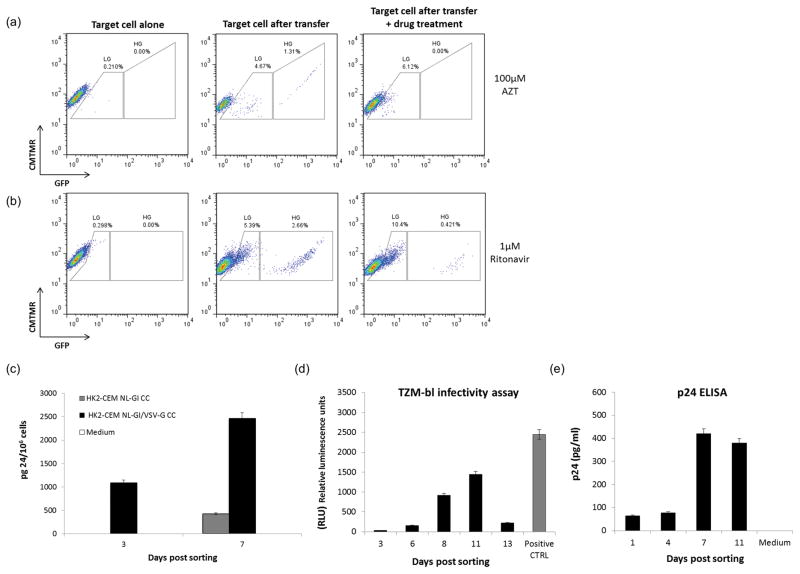
Figure 2. Antiretroviral drug inhibition and viral particles release from infected RTE.
(a) HK2 cells were co-cultivated with infected CEM T cells in presence of 100 μM of AZT. (b) CEM T cells were pre-treated with 1 μM of Ritonavir before viral infection. Ritonavir was added again at the moment of the infection and throughout the co-culture with renal epithelial cells (c) GFP positive HK2 derived from a co-culture between T cells infected either with NL-GI (grey histogram) or with a VSV-G pseudotyped NL-GI (black histogram), were flow sorted and p24 measured in the cell supernatants. Supernatants were collected at day 3 and 7 post sorting (fresh medium was added to the cells after supernatant removal at day 3) and analyzed for p24 as described in Materials and Methods. The amount of p24 is expressed per 106 cells. (d) Supernatants from flow sorted GFP positive HK2 were collected at different time points and analyzed for their ability to infect TZM-bl cells as described in Material and Methods. e) p24 measurement in supernatants from flow sorted GFP positive HK2 over time. Representative of three independent experiments.

