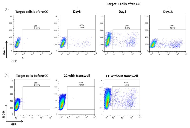
Figure 3. HIV-1 transfer from infected HK2 to uninfected CEM-SS T cells.
(a) Flow sorted GFP positive HK2 (day 6 post-sorting) were co-cultivated O.N. with uninfected CEM-SS T cells as described in Materials and Methods. At day 3 post co-culture CEM-SS T cells were analyzed for GFP expression by flow cytometry. Only the CMTMR-labeled population was gated for analysis. (b) Flow sorted GFP positive HK2 (day 4 post sorting) were co-cultivated O.N. with uninfected CEM-SS T cells in presence or absence of a transwell membrane (0.4μ pore size). At day 4 post co-culture, CEM-SS T cells were analyzed for GFP expression by flow cytometry. Representative of three independent experiments.

