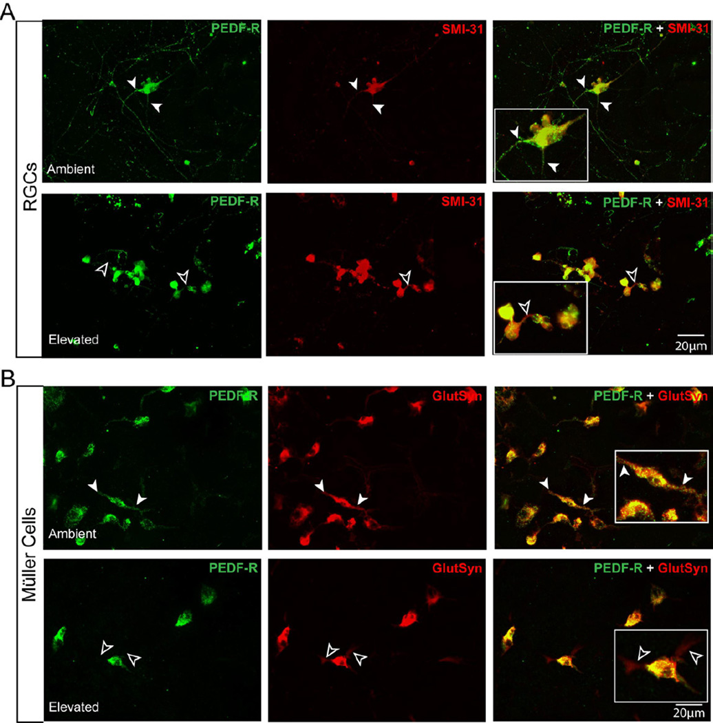
Figure 7.
Elevated pressure alters PEDF-R localization in RGCs and Müller cells in vitro. A. Fluorescent micrographs of PEDF-R (green) and SMI-31 (red) immunolabeling in purified, primary cultures of RGCs exposed to either ambient (top panels) or elevated (bottom panels) pressure for 48 hours reveals a reduction in localization of PEDF-R to neurites (black arrowheads) following exposure to elevated pressure, as compared to ambient pressure (white arrowheads). B. Fluorescent micrographs of PEDF-R (green) and glutamine synthetase (red) immunolabeling in purified, primary cultures of Müller cells exposed to either ambient (top panels) or elevated (bottom panels) pressure for 48 hours reveals a reduction in localization of PEDF-R to processes that is associated with process retraction (black arrowheads) following exposure to elevated pressure (black arrowheads) versus ambient pressure (white arrowheads).

