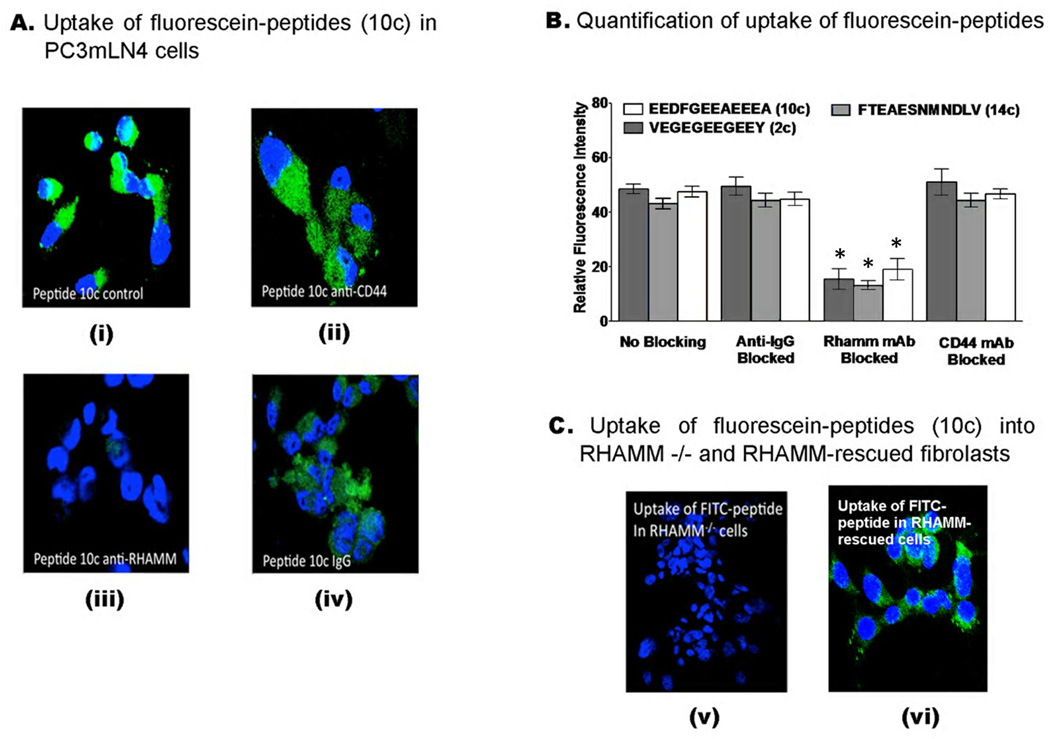
Figure 6. Uptake of FITC-peptides by PC3MLN4 prostate cancer cells.
(A) Confocal images of the uptake of FITC-conjugated peptide (10c) in prostate tumour cells using two-channel fluorescence confocal microscopy. Nuclei are shown in blue (DAPI) while FITC-conjugated peptides are shown in green. Prior to addition of dye-conjugated peptides, cells were incubated with non-immune IgG (iv), anti-RHAMM (iii) or anti-CD44 (ii) antibodies. Cells, which received no treatment (i) or were treated with non-immune IgG (iv) served as controls. A reduction in green channel fluorescence (FITC) was observed when cells are blocked with anti-RHAMM while no detectable decrease in FITC signal was observed for cells treated with anti-CD44 or IgG. (B) Uptake of 2c, 10c and 14c in PC3MLN4 tumor cells was quantified using ImageJ software, as described in Experimental Procedures. Values are mean fluorescence ± S.E.M Data were analyzed using one-way ANOVA. Significant differences (p < 0.05) are marked by asterisks. (C) FITC peptide (10c) uptake was not detectable in RHAMM−/− fibroblasts (v) but was observed when these cells were rescued by expressing a full length RHAMM cDNA (vi).

