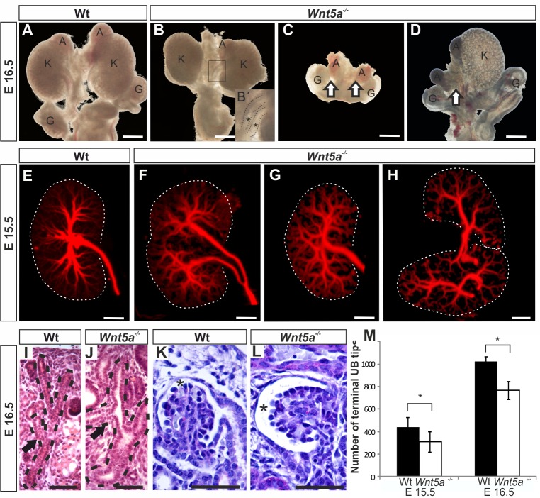
Fig 1. Wnt5a deficiency leads to severe metanephric kidney anomalies.
Embryonic urogenital systems (UGS) (A-D) and their kidneys (E-L) were dissected at E15.5 and E16.5. The UGS were inspected either as unstained (A-D) or their kidneys we subjected to Troma-1 antibody staining as whole mount and analysed with the optical projection tomography (OPT) to identify the pattern of the ureteric bud (UB) tree and the terminal UB derived tree tip counts (M) or sectioned (I-L). A) A normal urogenital system. The kidney (K), the gonad (G) and the adrenal gland (A) are marked. The Wnt5a deficiency leads to three categories of phenotypes; B) a kidney with duplex UB highlighted in the boxed image (the stars in B´), kidney hypoplasia (compare B with A), bilateral (C) or unilateral (D) renal failure. The OPT reveals variation noted in the pattern of UB tree development in the Wnt5a deficient embryonic kidneys when compared to control (compare F—H with E). The altered UB tree pattern can be depicted in the sections of the Wnt5a deficient kidneys when compared to control (compare J with I, dotted line, arrows). Wnt5a deficiency enlarges also the Bowman´s capsule lumen (asterisk) from control one (compare L with K, stars). Counting of the UB terminal tips from the OPT data indicates reduction in their number due to Wnt5a deficiency from controls M). Data in M are shown as means ± SD, n = 4–5 kidneys/group. Scale bars, A-D 400 μm, E-H 200 μm and I-L 50 μm. * P <0.05.

