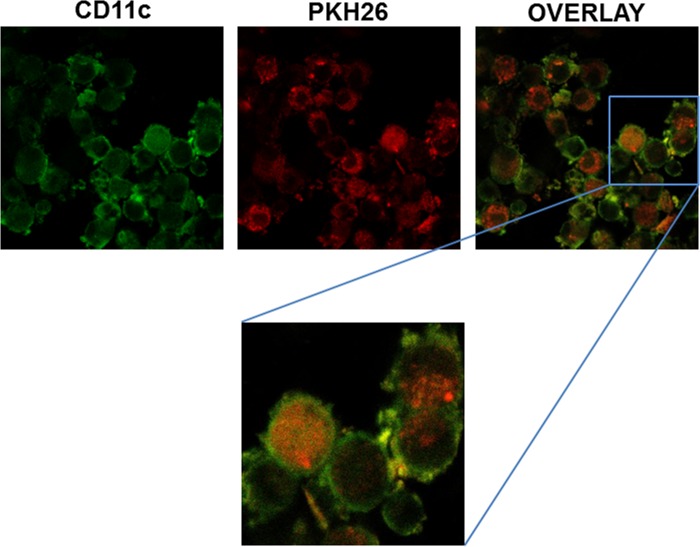
Fig 2. Verification of antigen load by confocal microscopy.
Confocal microscopy was used to verify cytosolic localization of tumour-derived antigens following 24 hour co-cultures of day-6 DCs and tumour cell lysate. Tumour masses were labelled with PKH26 Red Fluorescent Cell Linker (red) and DCs were stained with the monoclonal antibody anti-CD11c FITC (green). Antigen load is verified by visualization of tumour antigen (red) within dendritic cells (green). One representative of three independent experiments is shown; all images are 20X whereas zoom is 40X.

