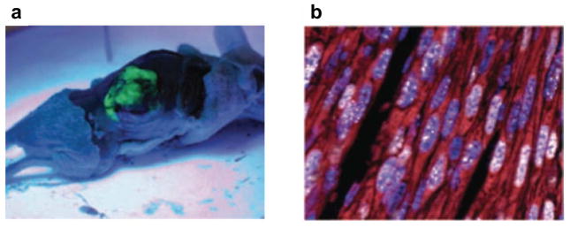
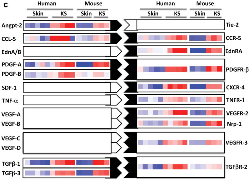
FIGURE 4. Mouse model of KSHV-induced KS.
a | Nude mice bearing enhanced green fluorescent protein-expressing tumours that are induced by subcutaneous injection of mouse endothelial cells transfected with Karposi’s sarcoma-associated herpesvirus (KSHV; also known as human herpesvirus 8 (HHV8)) KSHV Bac 36 (mECK36). b | Immunoflurorescence analysis of latency-associated nuclear antigen (LANA; white) and the Kaposi’s sarcoma (KS) marker podoplanin (red) in mECK36 tumours showing punctuated LANA staining that is characteristic of episomal KSHV in cell nucleus (DAPI Blue). c | Activation of paracrine and autocrine endothelial stimulation as shown by gene expression data (heat maps) from AIDS-KS41 and from the mECK36 mouse KS model171. Data are grouped by ligands (left) and receptors (right), which are present in both molecular signatures. Open connectors indicate paracrine stimulation by upregulation in KS of at least one of the receptor–ligand pairs; ligand and receptor closed connectors indicate upregulation of both receptor and ligand with potential for both paracine and autocrine stimulation. ANGPT2, angiopoietin 2; CCL5, chemokine, CC motif, ligand 5; CCR5, chemokine, CC motif receptor 5; CXCL12, chemokine CXC motif, ligand 12; EdnA, Endothelin A; NRP, neuropilin; PDGF, platelet-derived growth factor; TGFβ, transforming growth factor-β; TNF, tumour necrosis factor; VEGF, vascular endothelial growth factor.


