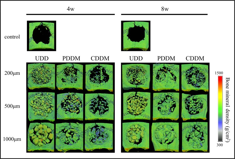
Fig 2. Radiographic analysis of specimens using μCT.
New bone formation around PDDM is enhanced in all particle sizes compared with CDDM in a time-dependent manner. The results of 1000 μm PDDM are especially noteworthy. On the contrary, almost all samples of UDD, with the exception of 1000 μm at 8 weeks, resulted in the defect being mostly occupied with dentin particles.

