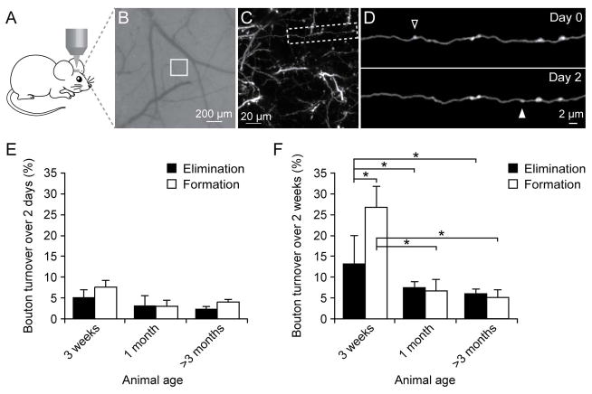
Figure 1. Axonal bouton turnover in young and adult barrel cortex.
(A) Transcranial two-photon imaging of the mouse barrel cortex. (B) CCD camera view of the vasculature of the barrel cortex below the thinned skull. The white box indicates the region where the two-photon image in (C) was obtained. (C) A low magnification image of dendrites and axons in Layer 1. A higher-magnification view of the axon segment in (C) is shown in (D). (D) Repeated imaging of an axonal branch over 2 days. The open and filled arrowheads indicate eliminated and newly formed axonal boutons over two days. (E) Elimination and formation rates of axonal boutons in three-week-old, one-month-old (young) and >3-month-old (adult) mice over a two-day interval. No significant differences were found in the elimination or formation rate among mice of various ages. (F) Elimination and formation rates of axonal boutons over two weeks in mice of various ages. Both the formation and elimination rates in 3-week-old mice were significantly higher than those in 1-month-old or adult mice. The formation rate of axonal boutons was significantly higher than the elimination rate at the age of 3 weeks. No significant difference in bouton formation or elimination rates was found over 2 weeks between 1-month-old and > 3-month-old mice. Data are presented as mean ± S.E.M. * P < 0.05.

