Fig. 3.
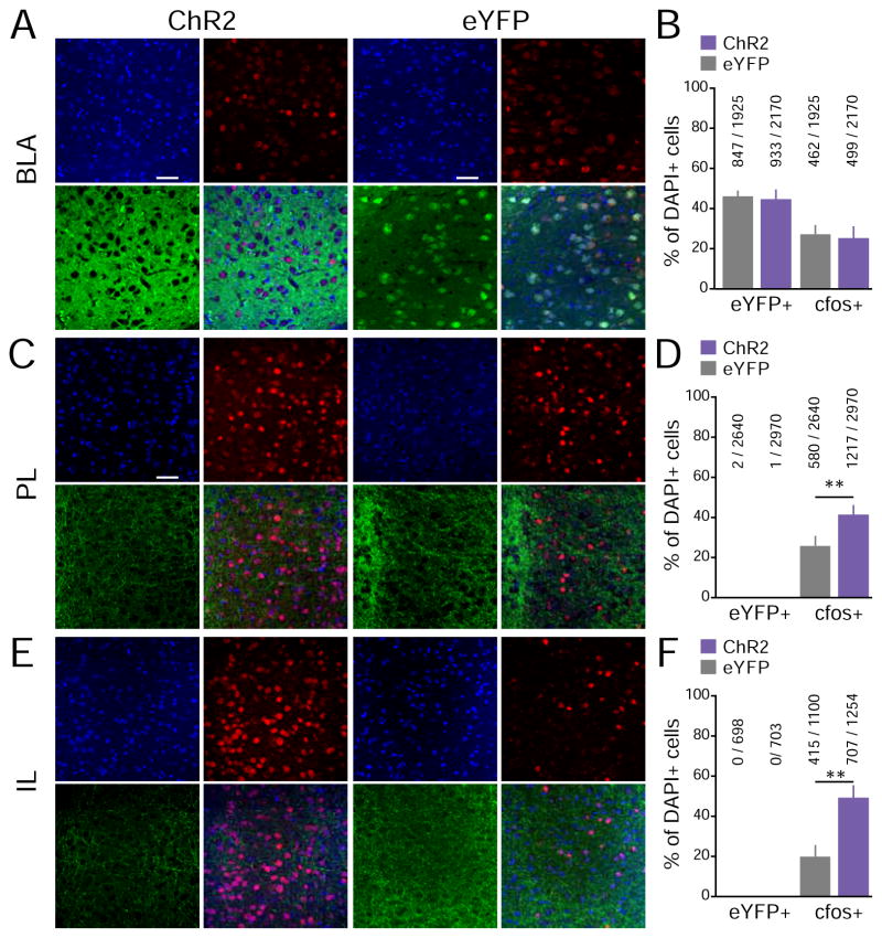
Activation of BLA inputs was sufficient to activate mPFC neurons. Mice underwent a 3-min photostimulation session in their homecage ~90 min prior to being sacrificed. Immunoreactivity to cfos was used as a proxy for neuronal activity. (ChR2 group n = 9, eYFP group n = 8). (A) 40X confocal images of the BLA from representative ChR2 and eYFP mice (blue: DAPI+ cells, green: eYFP+ cells, red: cfos+ cells). (B) Percentage of eYFP+ and cfos+ cells in the BLA, relative to the total DAPI+ cell counts. No significant differences were detected between the ChR2 and eYFP-control groups. (C) 40X images of PL sub-regions of the mPFC. (D) Percentage of eYFP+ and cfos+ cells in the PL sub-region of the mPFC, relative to DAPI counts. (E) 40X images of IL sub-regions of the mPFC. (D) Percentage of eYFP+ and cfos+ cells in the IL subr-region of the mPFC, relative to DAPI counts. As expected, almost no mPFC cell was eYFP+. The proportion of cfos+ mPFC (Both PL and IL) cells was significantly higher in the ChR2 group than the eYFP-control group, suggesting that photostimulation of BLA inputs facilitate mPFC activity.

