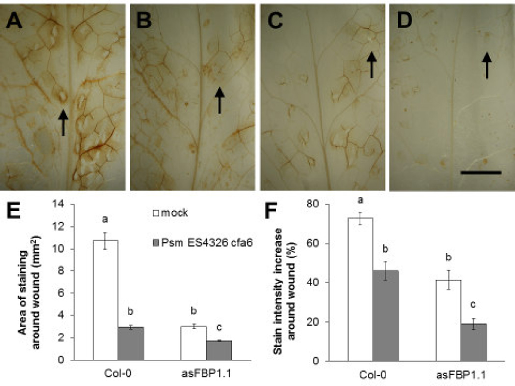
Fig. 5.
Infection with P. syringae reduces plant hydrogen peroxide levels upon S. flava feeding. A-D) Staining of hydrogen peroxide production (brown coloration) using 3,3’-diaminobenzidine tetrahydrochloride (DAB) in leaves of wild-type (Col-0) (A–B) and asFBP1.1 mutant (C–D) Arabidopsis plants that were either pre-infected with Psm ES4326 cfa6 four days prior to insect feeding (B and D) or mock-inoculated (A and C), left with adult female S. flava flies for 24 hours. Note the difference in the relative contrast in staining around the feeding sites (“wounds”) compared to the undamaged surrounding tissue between leaves pre-infected with Psm ES4326 cfa6 and mock-inoculated leaves, and the area of staining around individual feeding sites. Black arrows indicate sites of oviposition in which an egg was laid in the feeding site. E-F) Leaves representative for each treatment group, such as the leaves of which a close up is shown in (A–D), were selected for quantitative analysis. Letters in (E–F) indicate statistically significant differences (two-way ANOVA with post hoc Tukey’s HSD tests, P < 0.05). Error bars represent standard error of the mean. Scale bar in (D) for (A–D) indicates 5 mm.

