Figure 2.

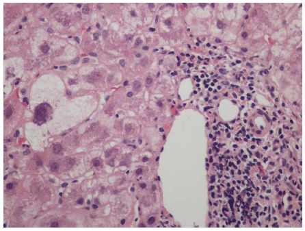
Lymphocyte predominant portal inflammation and a damaged bile duct (Poulsen Christofferson lesion). Periportal hepatocytes show ballooning/feathery degeneration. Hematoxylin and eosin stain, magnification × 200.

Lymphocyte predominant portal inflammation and a damaged bile duct (Poulsen Christofferson lesion). Periportal hepatocytes show ballooning/feathery degeneration. Hematoxylin and eosin stain, magnification × 200.