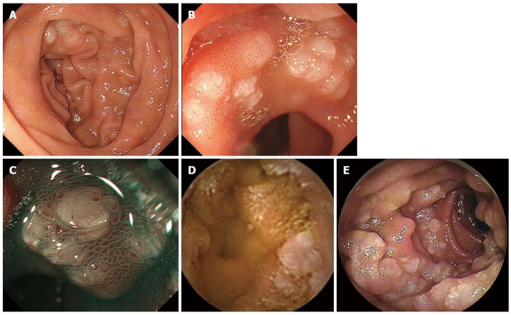
Figure 1.

Endoscopic features of intestinal follicular lymphoma in a 63-year-old woman. A: This case was diagnosed as follicular lymphoma with duodenal and jejunal lesions, and mesenteric lymph node involvement. The duodenal lesions are observed as multiple whitish nodules; B: Magnifying observation reveals opaque white depositions; C: Narrow-band imaging visualizes dilated microvessels on the surface of the white depositions; D: Video capsule enteroscopy shows multiple whitish granules in the jejunum; E: Double-balloon enteroscopy images of a jejunal lesion.
