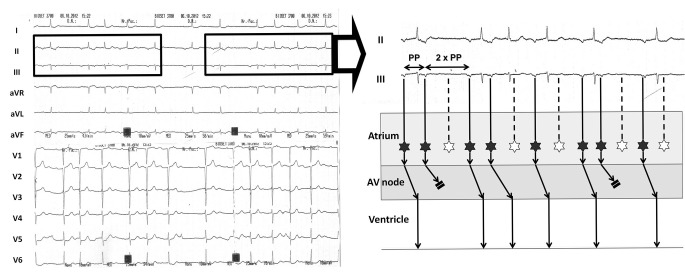
Fig. 1.
Left: Twelve-lead ECG at admission. The rhythm is regularly irregular, and there is a repetitive pattern in the rhythm (boxes); this suggests the presence of Wenckebach phenomena. Right: Ladder diagram depicting the mechanism of the arrhythmia. For simplicity, only leads II and III from the boxes on the left are displayed. The atrial rhythm is ectopic (P waves are negative in inferior leads) and irregular, with the longer PP interval twice the shorter PP interval. The atrial ectopic rate is 130 beats/min; hence, this is atrial tachycardia (originating from the inferior aspect of the atria) with variable Mobitz type II exit block to the atrium (in the ladder diagram, black stars denote atrial ectopic beats; white stars denote ectopic beats with exit block to the atrium). Furthermore, the atrial tachycardia is conducted to the ventricle with progressively longer PR intervals until some P waves are blocked. This behaviour is typical of Wenckebach phenomena in the atrioventricular (AV) node

