
FIGURE 2.
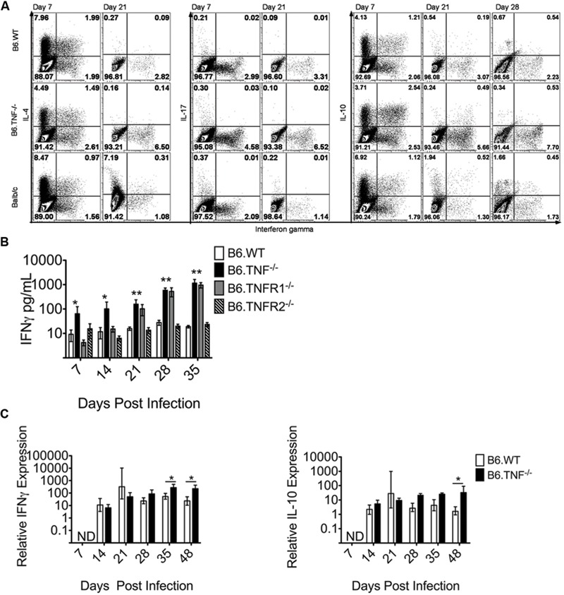
TNF-TNFR1 signaling pathway deficiency results in elevated IFN-γ expression after L. major infection. (A) Cytokine expression in CD4+ T cells from popliteal draining LNs of L. major infected Wt, Tnf-/- and BALB/c mice was analyzed. The use of the Tnf-/- mice constitutes the disruption of the complete TNF-TNFR1 signaling pathway. Intracellular flow cytometry was used to determine the proportional expression of IFN-γ, IL-4, IL-17, and IL-10 in CD4+ T cells at day 7 and 21 (and in the case of IL-10 also day 28) p. i. The experiment was performed two times independently and a representative staining is shown. (B) The concentration of IFN-γ in the serum of L. major infected Wt, Tnf-/-, Tnfr1-/-, and Tnfr2-/- was determined. The data are presented as mean (±SEM; n = 5–6 per genotype). Results are representative of at least 3 independent experiments. (C) The relative expression of IFN-γ and IL-10 in the footpad lesion of infected Wt or Tnf-/- mice was compared to uninfected controls. Relative expression was calculated relative to β-actin as described (Meissner et al., 2003). The data are presented as median (±SEM; n = 3–5 mice, representative of two independent experiments). The white bar represents Wt, the black bar Tnf-/- mice. The non-parametric Mann–Whitney U-test was used for analysis of experiments displayed in (B,C) to test for statistical differences (∗p < 0.05 ∗∗p < 0.01).