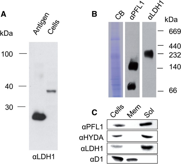
Fig. 1.

Immunochemical analysis of size and location of Cr-LDH1. (A) Immunoblot analysis of total C. reinhardtii cell extract (8.5 × 10⁵ cells loaded) using Cr-LDH1-specific antibodies (αLDH1) with approximately 10 ng of E. coli-expressed LDH1 antigen as control. (B) Analysis of Cr-LDH1 oligomeric state in whole-cell extracts separated by 1D BN-PAGE and probed with Cr-LDH1 antibodies (αLDH1). A PFL1 immunoblot (αPFL1) is provided for comparison, and loading is indicated by a Coomassie Brilliant Blue (CB)-stained gel. (C) Immunodetection of Cr-LDH1 in whole-cell extract (Cells) and in membrane (Mem) and soluble (Sol) fractions. Approximately 40 µg of protein was loaded per lane. HYDA, PFL1 and the D1 subunit of PSII were used as controls.
