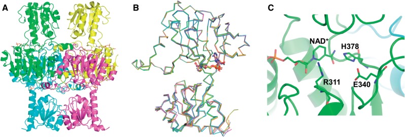
Fig. 2.
Crystal structure of His-tagged Cr-LDH1 (PDB:4ZGS). (A) Ribbon diagrams of the four chains proposed to form the tetramer. (B) Superposition of each of the four chains. NAD+ is shown in stick form. (C) View of NAD+ and the conserved histidine (H378), arginine (R311) and glutamate (E340) residues in the active site, shown in stick form. Numbering of residues is based on the decoded gene sequence (Supplementary Fig. S7). For the deposited structure, these residues are equivalent to H335, R268 and E297. Images were constructed using Pymol software.

