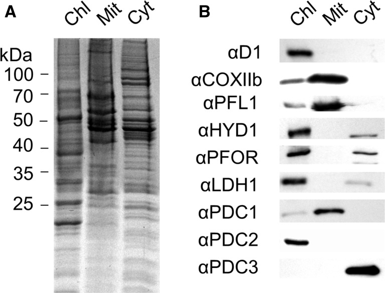
Fig. 4.
Determination of the subcellular distribution of pyruvate-degrading and fermentative enzymes in C. reinhardtii. Analysis of purified chloroplastic (Chl), mitochondrial (Mit) and cytoplasmic (Cyt) fractions, showing (A) a Coomassie Brilliant Blue (CB)-stained gel, (B) immunoblots. An 8 µg aliquot of protein was loaded per lane.

