Figure 2.
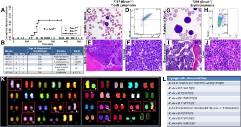
Brca1 deficiency increases susceptibility of mice to HMs characterized by leukemic infiltration of multiple organs consistent with the Bethesda criteria.14,15 (A) Cumulative disease incidence curves for Brca1+/+ (dash-dot-dot line), Brca1+/− (dashed line), and Brca1−/− (solid line) mice. Statistical significance was calculated by using the log-rank test. The number of mice in each cohort at each time point analyzed is listed in supplemental Figure 9H. (B) Characteristics of diseased mice and specific HM diagnosis. Presence or absence of a Trp53 mutation in each tumor is indicated. (C-F) Organs isolated from 7167, a Brca1−/−mouse that developed a T-cell lymphoma. (C) PB smear with Wright-Giemsa stain showing the presence of lymphoma cells (magnification ×10). (D) PB flow cytometry analysis using antibodies against T-lymphoid markers CD4 and CD8, showing the malignant cells to be CD4+/CD8+. (E) BM stained with hematoxylin and eosin (H&E), showing extensive involvement with lymphoma (magnification ×10). (F) Thymus stained with H&E, showing effacement of the normal thymic architecture by infiltrating lymphoma cells (magnification ×50). (G-J) Organs isolated from B7258, a Brca1−/− mouse that developed an erythroleukemia. (G) PB smear with Wright-Giemsa stain showing many erythroid blasts (magnification ×50). (H) PB flow cytometry analysis using antibodies against CD71 and Ter119. (I) BM stained with H&E, showing extensive leukemic involvement (magnification ×10). (J) Liver stained with H&E, showing extensive infiltration by leukemic cells (magnification ×50). (K) Spectral karyotyping analysis of erythroleukemia cells from a secondary transplant recipient mouse revealed an abnormal clone characterized by structural rearrangements: karyotype: 40,XX,t(1;15)(D;E), t(12;17)(D3;E2), and del(14)(D3E3). (L) Cytogenetic abnormalities identified within the erythroleukemia. del, deletion; idem, the same as the stemline clone listed first.

