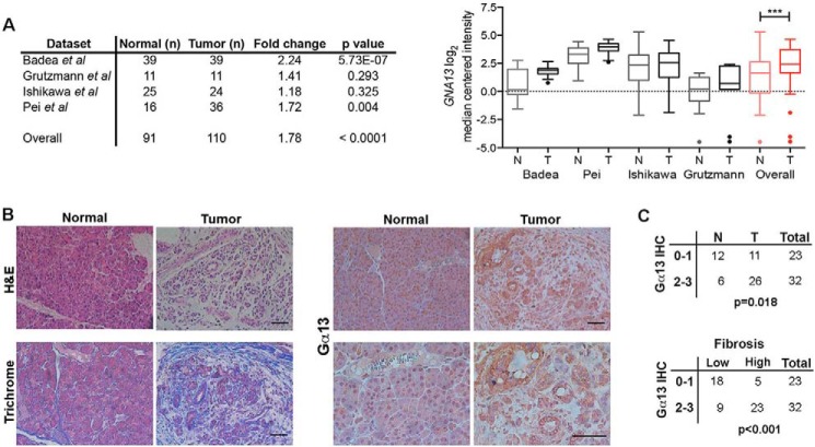
FIGURE 1.
Gα13 is up-regulated in human PDAC tumors. A, the relative expression of GNA13 mRNA was evaluated in human PDAC tumors (T) relative to normal (N) pancreas using the publicly available Oncomine database. Only databases that contained at least 10 normal specimens were analyzed. Overall statistical analyses of Oncomine data were performed using Student's t test. ***, p < 0.001. B and C, human pancreatic TMAs generated by the Pathology Core Facility at Northwestern University or purchased from Protein Biotechnologies were H&E-stained, trichrome-stained (blue = fibrosis), or immunostained with anti-Gα13 antibody (B). The relative staining was graded as low (0 or 1+) or high (2+ or 3+) as detailed under “Experimental Procedures.” The relative expression of Gα13 in tumor tissue and adjacent normal was assessed by Fisher's exact test (C). The extent of fibrosis was determined by the blue trichrome stain in the TMA specimens: <25% of the TMA core with blue stain (low) or >25% of the TMA core with blue stain (high). The relationship between Gα13 expression in the tissue samples and the extent of fibrosis was assessed by Fisher's exact test (C). IHC, immunohistochemistry. Scale bars, 50 μm.

