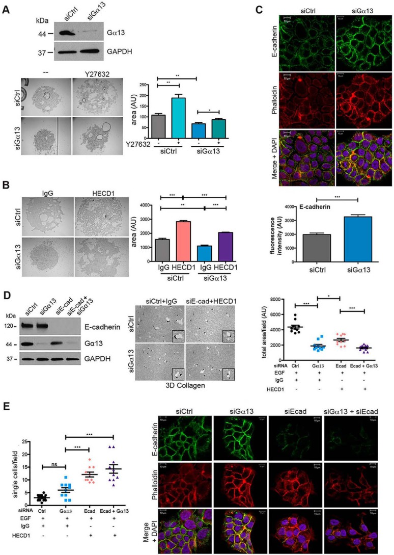
FIGURE 5.
Gα13 knockdown enhances E-cadherin-mediated cell-cell adhesion, but E-cadherin knockdown fails to reverse the effect of Gα13 siRNA on proteolytic invasion. A, CD18-tet-MT cells were transfected with siRNA for 96 h and then trypsinized and resuspended (2.5 × 10⁶ cells/ml of DMEM + 10% FBS +doxycycline) in the presence or absence of Y27632 (10 μm). A 10-μl drop of the cell suspension was hung on the undersurface of a tissue culture lid, incubated overnight at 37 °C, and photographed the next morning. The area covered by the cluster of cells was measured using ImageJ. B, CD18-tet-MT cells were transfected with siRNA for 96 h and then trypsinized and processed for the hanging drop assay in the presence of IgG or the function-blocking E-cadherin antibody HECD1. Hanging drops were incubated overnight at 37 °C and photographed the next morning. The area covered by the cluster of cells was measured using ImageJ. C, CD18-tet-MT cells were plated on glass coverslips, transfected with siRNA against Gα13, grown in the presence of doxycycline for 48 h, and fixed with 4% paraformaldehyde and permeabilized with 0.1% TritonX-100. Cells were stained for E-cadherin and phalloidin and imaged using a Zeiss Axiovert LSM510 Meta confocal microscope. The relative fluorescence intensity at cell-cell junctions was quantified using ImageJ. D and E, CD18-tet-MT cells were transfected with siRNA against Gα13, E-cadherin, or both Gα13 and E-cadherin. The effect on E-cadherin and Gα13 knockdown was determined by Western blotting. Prior to plating cells in three-dimensional (3D) collagen, cells were incubated with IgG or the E-cadherin (Ecad) function-blocking antibody HECD1. Cells were grown in three-dimensional collagen for 72 h with doxycycline in the presence of EGF (20 ng/ml). Gels were then processed to examine the effect on invasion as detailed in Fig. 2, and relative invasion was quantified (D). The number of single cells present in three-dimensional collagen per field in the invasion assay was quantified (E, left panel). CD18-tet-MT cells were plated on glass coverslips, transfected with siRNA against Gα13 and/or E-cadherin, grown in the presence of doxycycline for 48 h, fixed, permeabilized, and stained for E-cadherin and phalloidin (E, right panel). The statistics of the invasion assay were performed using one-way ANOVA followed by Dunnett's post-test. The statistics of the hanging drop assay were performed using Student's t test. ns, not significant; *, p < 0.05; **, p < 0.01; ***, p < 0.001. The results are representative of four independent experiments. Ctrl, control.

