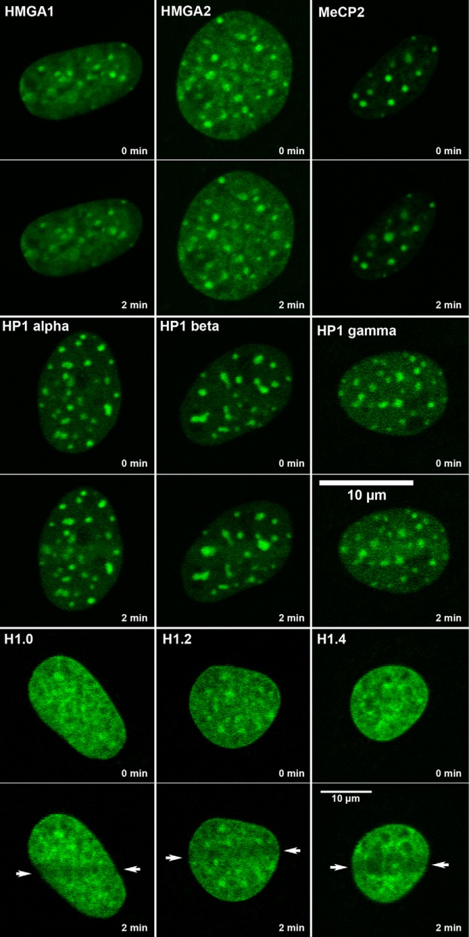
FIGURE 3.
Lack of displacement of non-histone heterochromatin proteins at sites of DNA damage. MEFs were transfected with GFP-tagged versions of HMGA1, HMGA2, MeCP2, and the three isoforms of HP1. DNA damage was introduced by laser microirradiation and the dynamics of these proteins were followed by time lapse microscopy. The figure shows images collected immediately before and ∼2 min after the introduction of DNA damage. The lower panel shows the displacement of example histone H1 variants after laser microirradiation.

